- Proteins are biological polymers composed of amino acids.
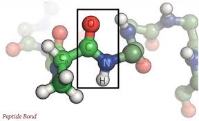
- Amino acids linked together by peptide bond to form a polypeptide chain.
- One or more polypeptide chain twisted into 3D shape to form protein.
- Protein complex shapes includes various folds, loops and curves.
- It is an essential constituent of all cells.
- It is in every part of body, skin, muscles, hair, blood, organs, eyes, finger nails and bones.
- These are macromolecular polymers composed of amino acid as basic unit.

- Protein molecules: fibrous: elongated and insoluble. And Globular: compact, soluble, spherical.
| Globular protein | Fibrous protein |
| Polypeptide chains are compactly folded to form spherical or globular shape | Polypeptide chains are extended along one axis and are spherically wound to form fibers |
| 4 types of bonds: H-bonds, ionic bonds, disulphide bonds and hydrophobic bonds, which maintains a tertiary structure. | H-bonds between amino acids residue, so usually have high degree of secondary structure. |
| Soluble in water | Insoluble in water |
| Non-contractile | Contractile |
| Ex. Egg albumin, globulins, Hb, all enzymes, etc. | Ex. Alpha-keratin of hair, nails, claws, horns, etc. Elastin, Collagen |
| Long, parallel chains form fibres | |

Types of protein:
Simple protein:
Gives only amino acid on hydrolysis. Ex. Ribonuclease
Composed of only alpha-amino acids.
- Albumins: soluble in water and dilute in salt solution. It is present in egg white portion and in blood. It is neutral.
- Globulins: insoluble in water but soluble in dilute salt solution. It is present in antibodies in blood serum and as blood fibrinogen. It is neutral.
- Histones: soluble in water and insoluble in dilute ammonium hydroxide. It is basic in nature. Ex. Chromatin.
Conjugated protein:
Gives amino acid and non- amino acids components on hydrolysis.
Conjugated proteins:
| Class | Prosthetic group | Examples |
| Lipoprotein | Lipids | Beta1- lipoprotein of blood, yolk, serum, milk, and cell membranes |
| Glycoprotein | Carbohydrate | Immunoglobulin G |
| Phosphoprotein | Phosphate group | Casein of milk |
| Hemoprotein | Heme (Fe porphyrin) | Haemoglobin |
| Flavoprotein | Flavin nucleotide | Succinate dehydrogenase |
| Metalloprotein | Fe Zn Ca Mo Cu | Ferritin Alcohol dehydrogenase Calmodulin (any Ca-bindingg protein) Dinitrogenase Plastocyanin |
- Prosthetic group: an inorganic/organic component (non amino acid) that is covalently bound to a protein and essential for its activity.
- Co–factor: an inorganic/organic component is not covalently bound to a protein.
Functional diversity of proteins:
- Enzymes: Hexokinase, kinase, etc.
- Transport proteins: haemoglobin, lipoprotein, membrane transport proteins.
- Nutrient and storage proteins: albumin, casein.
- Contractile protein: actin, myosin, tubulin, etc.
- Structural protein: collagen, desmosine, elastin, keratin, spider web protein.
- Defense protein: immunoglobulin, fibrinogen, thrombin, snake venom, bacterial toxins, rcin, abrin, etc.
- Regulatory protein: hormones, GTP – binding protein.
- Other proteins – antifreeze protein, monellin, etc.
Homologous proteins
Group of protein perform same function in all organism but are evolutionary related (structural resemble)
Example: Hb – oxygen transport
Mb – tissues O2 – transport
Cytochrome – C
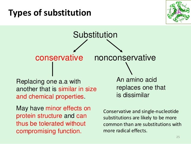
Conservative substitution:
When one amino acid of one part is changed by another. Ex: 1 polar acid changed by 1 another polar amino acid.
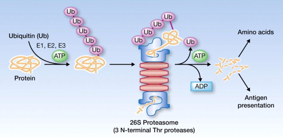
Ubiquitin
With help of ubiquitin, protein degrade after the completion of its function, if ubiquitin do not function then a cell gets block.
Protein conformation
It is stabilized by weak interactions and the stability of unfold state of protein is maintained by high degree of entropy and H-bonding interaction of many group in polypeptide chain.
Conformation:
Spatial arrangement of atoms in a protein and a change in conformation could occur by rotation about single bond.
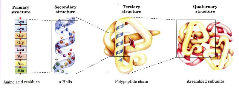
Levels of architecture of proteins:
- Primary structure: it refers to amino acid sequence and all covalent bond between amino acids and location of disulphide bonds.
- Secondary structure: refers to regular recurring arrangement.
- Tertiary structure: refers to spatial relationship between all amino acids.
- Quaternary structure: refers too spatial relationship between different polypeptides.

Primary structure
It is sequence of amino acids in polypeptide chain.
Unique order in which amino acids are linked together to form a protein.
There is the peptide linkage in the form of bonding.
Structure of peptides are: amino acids whose amino group is free and group called N-terminal end, and other whose carboxyl group is free and group called C-terminal end.

Proteins are constructed from a set of 20 amino acids. Generally, amino acids have following structural properties:
C (alpha-C) bonded to 4 group below:-
- H – atom
- -NH2 group
- -COOH group
- Variable or R group
Amino acids sequence of a protein is determined by information found in a cellular genetic code.
Secondary structure
Formed by folding of primary structure.
2 polypeptide are held together by H-bonds.
The coiling or folding of a polypeptide chain that gives proteins its 3D structure.
It is the arrangement od amino acids chain due to the H-bonds between atoms at different parts of the chain.
Ex: collagen and silk – fibrin
Proteins can assume 2 conformational structure: α-helix and β-pleated sheet
α-helix:
- Resembles a coiling spring
- Secured by H-bonding in polypeptide chain.
- Constraints which affect the stability of α-helix:
- Electrostatic repulsions (or attraction) between amino acid residues with charged R-group.
- The bulkiness of adjacent R-group.
- The interaction between amino acid side chain spaced 3 or 4 residues apart.
- Occurrence of proline structure.
- Interaction between amino acid at ends of helix and electric dipole inherent to this structure.
- There is intrachain H-bonding.
Β-pleated sheet:
- Appears to be folded or pleated and is held together by H-bonding between polypeptide units of the folded chain that lie adjacent to one another.
- 2 types:
- Parallel beta sheets:-chains of polypeptides, which run in the same direction.
- Anti-parallel beta sheets:-chains of polypeptides, which run in the opposite direction to eachother.
- These are both intra and inter- chain H bonding, it is extended or zig-zag conformations.

| Α-helix | Β– helix |
| Helical conformation | Zig-Zag conformation |
| Interchain | Inter and Intra- chain |
| R-group project outside | R-group is projected alternately |
Tertiary structure
- Refers to the 3D-structure of polypeptide chains of a protein.
- There are several types of bonds and forces that hold a protein in its tertiary structure.
- Hydrophobic interactions: Greatly contribute to folding and shaping of a protein. ‘R’ group – amino acid: hydrophobic or hydrophilic.
- Hydrophilic R-group: seek contact with aqueous environment
- Hydrophobic R-group: seek to avoid water and position themselves towards the center of protein.
- Hydrogen bonding: In the polypeptide chain and between amino acid R group helps to stabilize protein structure by holding the protein in shape established by hydrophobic interactions.
- Due to protein folding, Ionic bonding can occur between positively and negatively charged R group that come in close contact with one another.
- Folding also results in Covalent bonding between R group of cysteine amino acid. This type of bonding forms: disulfide bridge.
- Interactions called Vander wall forces also assist in stabilization. These contribute to bonding that occurs between molecules.

Quaternary structure
- Proteins are formed by more than one polypeptide chain and joined together by covalent bond.
- Each polypeptide is called as a subunit.
- Quaternary structure formed by interachain.
- When protein formed by similar subunit : homogenous quaternary structure, and when protein formed by dissimilar subunit: heterogenous quaternary structure. It is consists of 2 identical α-chain and 2- identical β-promoter.
- Most stable structure.

Stability
Less stable to more stable:
Primary to secondary to tertiary to quaternary
Chemical bonds involved in protein structure
- Strong bonds
- Peptide bonds: covalent bonds formed by dehydration synthesis between alpha-carboxyl group of one amino acid and alpha-amino group of adjacent amino acid.
- Disulphide bonds: covalent bonds formed between the S-containing cysteine residue of polypeptide chain.
- Weak bonds
- Hydrogen bonds: sharing of H-atoms between the –N and –C=O of different peptide bond.
- Hydrophobic bonds: when two chains of amino acids come together, no true bonds are formed.
- Ionic, electrostatic or salt linkages: formed between closely lying –COO and –NH3 group of different amino group residues.


Oneplus Nord CE4 (Dark Chrome, 8GB RAM, 256GB Storage)
₹26,999.00 (as of April 19, 2024 08:30 GMT +00:00 - More infoProduct prices and availability are accurate as of the date/time indicated and are subject to change. Any price and availability information displayed on [relevant Amazon Site(s), as applicable] at the time of purchase will apply to the purchase of this product.)
iQOO Z9 5G (Brushed Green, 8GB RAM, 256GB Storage) | Dimensity 7200 5G Processor | Sony IMX882 OIS Camera | 120Hz AMOLED with 1800 nits Local Peak Brightness | 44W Charger in The Box
₹21,999.00 (as of April 19, 2024 08:30 GMT +00:00 - More infoProduct prices and availability are accurate as of the date/time indicated and are subject to change. Any price and availability information displayed on [relevant Amazon Site(s), as applicable] at the time of purchase will apply to the purchase of this product.)
Redmi 13C 5G (Startrail Silver, 4GB RAM, 128GB Storage) | MediaTek Dimensity 6100+ 5G | 90Hz Display
₹10,499.00 (as of April 19, 2024 08:30 GMT +00:00 - More infoProduct prices and availability are accurate as of the date/time indicated and are subject to change. Any price and availability information displayed on [relevant Amazon Site(s), as applicable] at the time of purchase will apply to the purchase of this product.)
Redmi 13C 5G (Starlight Black, 4GB RAM, 128GB Storage) | MediaTek Dimensity 6100+ 5G | 90Hz Display
₹10,499.00 (as of April 19, 2024 08:30 GMT +00:00 - More infoProduct prices and availability are accurate as of the date/time indicated and are subject to change. Any price and availability information displayed on [relevant Amazon Site(s), as applicable] at the time of purchase will apply to the purchase of this product.)
iQOO Z9 5G (Brushed Green, 8GB RAM, 128GB Storage) | Dimensity 7200 5G Processor | Sony IMX882 OIS Camera | 120Hz AMOLED with 1800 nits Local Peak Brightness | 44W Charger in The Box
₹19,999.00 (as of April 19, 2024 08:30 GMT +00:00 - More infoProduct prices and availability are accurate as of the date/time indicated and are subject to change. Any price and availability information displayed on [relevant Amazon Site(s), as applicable] at the time of purchase will apply to the purchase of this product.)
Redmi 13C (Stardust Black, 6GB RAM, 128GB Storage) | Powered by 4G MediaTek Helio G85 | 90Hz Display | 50MP AI Triple Camera
₹8,699.00 (as of April 19, 2024 08:30 GMT +00:00 - More infoProduct prices and availability are accurate as of the date/time indicated and are subject to change. Any price and availability information displayed on [relevant Amazon Site(s), as applicable] at the time of purchase will apply to the purchase of this product.)
Oneplus Nord CE4 (Dark Chrome, 8GB RAM, 128GB Storage)
₹24,999.00 (as of April 19, 2024 08:30 GMT +00:00 - More infoProduct prices and availability are accurate as of the date/time indicated and are subject to change. Any price and availability information displayed on [relevant Amazon Site(s), as applicable] at the time of purchase will apply to the purchase of this product.)
Redmi 13C 5G (Startrail Green, 4GB RAM, 128GB Storage) | MediaTek Dimensity 6100+ 5G | 90Hz Display
₹10,499.00 (as of April 19, 2024 08:30 GMT +00:00 - More infoProduct prices and availability are accurate as of the date/time indicated and are subject to change. Any price and availability information displayed on [relevant Amazon Site(s), as applicable] at the time of purchase will apply to the purchase of this product.)
Redmi 13C (Starfrost White, 6GB RAM, 128GB Storage) | Powered by 4G MediaTek Helio G85 | 90Hz Display | 50MP AI Triple Camera
₹8,699.00 (as of April 19, 2024 08:30 GMT +00:00 - More infoProduct prices and availability are accurate as of the date/time indicated and are subject to change. Any price and availability information displayed on [relevant Amazon Site(s), as applicable] at the time of purchase will apply to the purchase of this product.)
realme 12 Pro 5G (Submarine Blue, 8GB RAM 256 GB Storage)
₹23,378.00 (as of April 19, 2024 08:30 GMT +00:00 - More infoProduct prices and availability are accurate as of the date/time indicated and are subject to change. Any price and availability information displayed on [relevant Amazon Site(s), as applicable] at the time of purchase will apply to the purchase of this product.)
Amazon Brand - Jam & Honey Penguin, Plush/Soft Toy for Boys, Girls and Kids, Super-Soft, Safe, Great Birthday Gift (Black and White, 17 cm)
₹169.00 (as of April 19, 2024 08:30 GMT +00:00 - More infoProduct prices and availability are accurate as of the date/time indicated and are subject to change. Any price and availability information displayed on [relevant Amazon Site(s), as applicable] at the time of purchase will apply to the purchase of this product.)
Kathuzz Non Scratch Dish Wash Cloth (5 Pack) Two Layer Mesh Wire Cloth for Kitchen Reusable Non Scratch Wire Dish Cloth Multipurpose Wire Dishwashing Rags for Wet and Dry Cleaning Scrubber Dish Cloth
₹398.00 (as of April 19, 2024 08:30 GMT +00:00 - More infoProduct prices and availability are accurate as of the date/time indicated and are subject to change. Any price and availability information displayed on [relevant Amazon Site(s), as applicable] at the time of purchase will apply to the purchase of this product.)
rts Universal Travel Adapter, International All in One Worldwide Travel Adapter and Wall Charger with USB Ports with Multi Type Power Outlet USB 2.1A,100-250 Voltage Travel Charger (Black)
₹587.00 (as of April 19, 2024 08:30 GMT +00:00 - More infoProduct prices and availability are accurate as of the date/time indicated and are subject to change. Any price and availability information displayed on [relevant Amazon Site(s), as applicable] at the time of purchase will apply to the purchase of this product.)
The Derma Co Hyaluronic Sunscreen Aqua Ultra Light Gel With Spf 50 Pa++++ For Broad Spectrum, UV A, UV B & Blue Light Protection For Oily Skin - 50G(Dermaco), Pack Of 1
₹448.00 (as of April 19, 2024 08:30 GMT +00:00 - More infoProduct prices and availability are accurate as of the date/time indicated and are subject to change. Any price and availability information displayed on [relevant Amazon Site(s), as applicable] at the time of purchase will apply to the purchase of this product.)
Aqualogica Glow+ Dewy Sunscreen SPF 50 PA++++ | UVA/B & Blue Light Protection for Men & Women | Oily, Dry, Sensitive & Combination Skin | Fragrance-Free | 50g
₹394.00 (as of April 19, 2024 08:30 GMT +00:00 - More infoProduct prices and availability are accurate as of the date/time indicated and are subject to change. Any price and availability information displayed on [relevant Amazon Site(s), as applicable] at the time of purchase will apply to the purchase of this product.)
Double layer Soap Dispenser for Bathroom Accessories Dishwasher Liquid Holder Liquid Dispenser Pump with Sponge Holder Kitchen Sink Accessories Items(multi colour)
₹169.00 (as of April 19, 2024 08:30 GMT +00:00 - More infoProduct prices and availability are accurate as of the date/time indicated and are subject to change. Any price and availability information displayed on [relevant Amazon Site(s), as applicable] at the time of purchase will apply to the purchase of this product.)
Lymio Men Cargo || Men Cargo Pants || Men Cargo Pants Cotton || Cargos for Men (Cargo-05-08)
₹699.00 (as of April 19, 2024 08:30 GMT +00:00 - More infoProduct prices and availability are accurate as of the date/time indicated and are subject to change. Any price and availability information displayed on [relevant Amazon Site(s), as applicable] at the time of purchase will apply to the purchase of this product.)
Allen Solly Men's Regular Fit Polo
₹550.00 (as of April 19, 2024 08:30 GMT +00:00 - More infoProduct prices and availability are accurate as of the date/time indicated and are subject to change. Any price and availability information displayed on [relevant Amazon Site(s), as applicable] at the time of purchase will apply to the purchase of this product.)
BSB HOME Microfiber 144 TC Aspire 2.O Collections Soft Breathable Wrinklefree Floral Printed Double Bedsheets with 2 Regular Size Pillow Covers, Color White Green Pink Rose
₹199.00 (as of April 19, 2024 08:30 GMT +00:00 - More infoProduct prices and availability are accurate as of the date/time indicated and are subject to change. Any price and availability information displayed on [relevant Amazon Site(s), as applicable] at the time of purchase will apply to the purchase of this product.)